Regenerative Medicine 2026: How Stem Cells and Bioengineering Are Rebuilding the Human Body
iPS cell therapy for Parkinson's involves reprogramming donor cells, differentiating them into dopamine-producing neurons, and transplanting them into the patient's brain
Introduction – Why This Matters
In my experience as a medical science writer who has spent years covering the frontier of regenerative medicine, I’ve witnessed a field transform from laboratory curiosity to clinical reality. I remember attending conferences a decade ago where researchers presented elegant experiments in petri dishes and mouse models, always concluding with the same refrain: “Translation to humans is still years away.” Those years have now arrived.
What I’ve found is that 2026 marks a watershed moment for regenerative medicine. The past eighteen months have delivered what many thought impossible: the world’s first regulatory approvals for induced pluripotent stem cell (iPS cell) therapies. Japan’s health ministry has green-lit treatments for Parkinson’s disease and severe heart failure, with products expected to reach patients this summer. A UC Davis team has safely performed the first-ever in-utero stem cell therapy for spina bifida, publishing results in The Lancet. Researchers at Cedars-Sinai have uncovered how transplanted neural stem cells preserve vision in degenerative eye disease, using single-cell analysis to reveal mechanisms that could lead to more powerful treatments.
These are not incremental advances. They represent a fundamental shift in how we approach disease—moving from managing symptoms to rebuilding damaged tissues and organs. The convergence of stem cell biology, tissue engineering, gene editing, and materials science has created capabilities that seemed like science fiction just a generation ago.
The numbers driving this field are staggering. Over 100 million people worldwide live with conditions potentially addressable by regenerative medicine—heart failure, neurodegenerative diseases, type 1 diabetes, spinal cord injury, and countless others. The global regenerative medicine market, valued at approximately $40 billion in 2025, is projected to exceed $100 billion by 2030. But beyond the market size, what excites me is the human impact: patients with Parkinson’s disease who may regain motor function, children with spina bifida who may walk, and individuals with heart failure who may avoid transplantation.
This guide will walk you through everything you need to know about regenerative medicine in 2026—how different approaches work, what clinical advances have been achieved, what regulatory milestones have been reached, and where this field is heading. Whether you’re someone personally affected by a condition that regenerative medicine might address, a healthcare professional seeking to understand emerging options, or simply curious about the future of medicine, this article will give you a comprehensive, practical understanding of regenerative medicine today.
Background / Context
The Promise of Regeneration
Regenerative medicine encompasses approaches that repair, replace, or regenerate damaged cells, tissues, and organs. Unlike conventional treatments that manage symptoms or slow disease progression, regenerative medicine aims to restore normal function by rebuilding what’s been lost.
The concept is ancient in its appeal—the dream of regenerating limbs like a salamander or healing wounds without scarring. But the scientific foundation emerged only in the past few decades, built on advances in stem cell biology, developmental biology, and materials science.
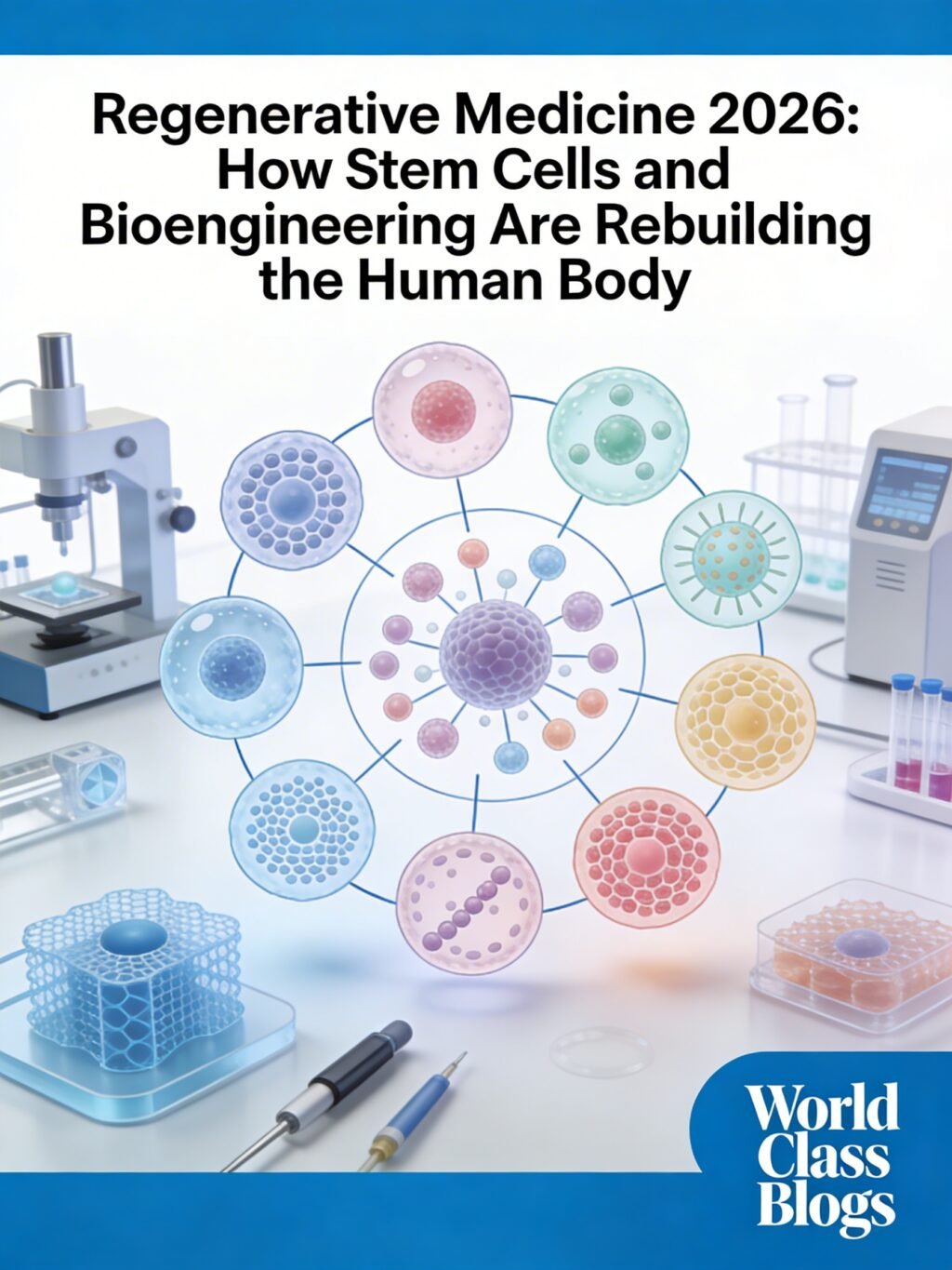
The Stem Cell Revolution
Stem cells are the raw material of regenerative medicine. Their defining properties—self-renewal and the ability to differentiate into specialized cell types—make them uniquely suited for tissue repair.
Embryonic stem cells (ESCs), first isolated in 1998, can become any cell type in the body. But their derivation from embryos raised ethical concerns that limited research funding and clinical translation in many countries.
Adult stem cells (also called somatic stem cells) are found in various tissues—bone marrow, fat, skin, and others. They’re more limited in differentiation potential but have been used clinically for decades. Hematopoietic stem cell transplantation for blood cancers is the oldest and most established stem cell therapy.
The game-changer came in 2006 when Shinya Yamanaka at Kyoto University discovered that introducing just four factors into adult skin cells could reprogram them into an embryonic-like state. These induced pluripotent stem cells (iPS cells) could become any cell type but were derived without embryos. Yamanaka received the Nobel Prize in 2012, and iPS cells have since become the workhorse of regenerative medicine research.
The Tissue Engineering Revolution
Stem cells alone aren’t enough—they need scaffolding, signals, and structure to form functional tissues. Tissue engineering combines cells with biomaterials and growth factors to create constructs that can be implanted or used to stimulate repair.
Early tissue engineering produced simple structures—skin grafts, cartilage patches. Today’s approaches are far more sophisticated, incorporating 3D printing, organ-on-a-chip technology, and even the ability to decellularize organs and repopulate them with patient cells.
The 2026 Landscape
As of 2026, regenerative medicine has achieved milestones that would have seemed impossible a decade ago:
Regulatory approvals: Japan has granted conditional approval to the world’s first iPS cell therapies—Amchepry for Parkinson’s disease and ReHeart for heart failure. These are not research products but commercially available treatments, marking the transition from experimental to clinical medicine.
Fetal therapy: UC Davis researchers have completed Phase 1 of the CuRe Trial, demonstrating that combining fetal surgery with placental-derived stem cells for spina bifida is safe. All six treated infants showed reversal of hindbrain herniation, and none required shunts for hydrocephalus .
Mechanistic understanding: Cedars-Sinai investigators have used single-cell analysis to reveal how neural stem cells preserve vision in retinitis pigmentosa—providing protective proteins, restoring retinal cells to a healthier state, reducing cellular stress, and maintaining retinal integrity .
Organ rejuvenation: Researchers are developing ex vivo machine perfusion platforms that could deliver rejuvenation therapies—including partial reprogramming—to donor organs before transplantation, potentially expanding the donor pool and improving outcomes.
Bioengineered tissues: Novel approaches to tissue clearing have enabled the creation of transparent decellularized human sclera as a corneal substitute, demonstrating anti-angiogenic and anti-fibrotic efficacy superior to donor corneas in preclinical models.
The field has also matured in its understanding of challenges. Safety concerns about tumor formation, immune rejection, and cell survival have been systematically addressed. Manufacturing processes have been scaled and standardized. Regulatory pathways have been established. Regenerative medicine is no longer a future promise—it’s a present reality.
Key Concepts Defined
Before diving deeper, let’s establish clear definitions of essential regenerative medicine terminology. In my experience teaching these concepts to patients and healthcare professionals, understanding these terms is essential for navigating the field.
Regenerative Medicine: A field of medicine that develops methods to regrow, repair, or replace damaged or diseased cells, tissues, and organs. It includes cell therapies, tissue engineering, gene editing, and other approaches aimed at restoring normal function.
Stem Cells: Undifferentiated cells capable of self-renewal and differentiation into specialized cell types. Different types have different potentials and sources.
Pluripotent Stem Cells: Cells that can differentiate into any cell type in the body. Includes embryonic stem cells and induced pluripotent stem cells.
Induced Pluripotent Stem Cells (iPS cells): Adult cells reprogrammed to an embryonic-like state by introducing specific factors (OCT4, SOX2, KLF4, cMYC). They can become any cell type without the ethical concerns of embryonic stem cells.
Mesenchymal Stem Cells (MSCs): Adult stem cells found in bone marrow, fat, and other tissues. They can differentiate into bone, cartilage, and fat cells and have potent immunomodulatory properties.
Hematopoietic Stem Cells (HSCs): Stem cells that give rise to all blood cell types. HSC transplantation (bone marrow transplant) is the oldest and most established stem cell therapy.
Tissue Engineering: An approach that combines cells, biomaterials (scaffolds), and bioactive molecules to create functional tissue constructs for implantation or for stimulating repair.
Biomaterials: Natural or synthetic materials used to create scaffolds that support cell attachment, growth, and organization. Examples include collagen, hyaluronic acid, synthetic polymers, and decellularized extracellular matrix.
Decellularization: The process of removing cells from a tissue or organ, leaving behind the extracellular matrix scaffold. This scaffold can then be repopulated with a patient’s own cells to create a personalized graft.
Ex Vivo Machine Perfusion: A technology that maintains organs outside the body by pumping oxygenated nutrient solution through their blood vessels. This creates a window for delivering therapies to improve organ quality before transplantation.
Partial Reprogramming: Transient expression of Yamanaka factors to reverse age-related cellular changes without erasing cell identity. This approach can restore youthful epigenetic signatures and enhance tissue function.
Yamanaka Factors: The four transcription factors (OCT4, SOX2, KLF4, cMYC) used to reprogram adult cells into induced pluripotent stem cells. Shinya Yamanaka’s discovery earned him the Nobel Prize in 2012.
Conditional and Time-Limited Approval: A regulatory pathway that allows marketing of promising therapies based on early clinical data, with requirements for continued data collection and periodic re-evaluation. Japan used this pathway for its first iPS cell approvals.
Hindbrain Herniation: A complication of spina bifida where the brainstem and cerebellum are pulled downward into the spinal canal. Reversal of hindbrain herniation is a key indicator of successful fetal surgery.
Retinitis Pigmentosa: A group of inherited eye diseases causing progressive vision loss due to degeneration of the light-sensitive retina. Stem cell approaches are being developed to preserve remaining vision.
How Regenerative Medicine Works (Step-by-Step Breakdown)
Understanding how regenerative medicine works requires looking at different approaches for different applications. Let me walk you through the major strategies and their step-by-step processes.
Approach 1: Cell Replacement Therapy (Parkinson’s Disease)
Parkinson’s disease involves progressive loss of dopamine-producing neurons in a specific brain region. Cell replacement aims to restore these neurons.
Step 1: Cell Source Generation: Healthy donor cells (typically from unrelated individuals) are reprogrammed into iPS cells. These pluripotent cells can be expanded indefinitely in culture.
Step 2: Directed Differentiation: The iPS cells are guided through developmental stages to become dopamine-producing neural precursor cells. This requires precise culture conditions, growth factors, and timing—typically weeks of carefully controlled differentiation.
Step 3: Quality Control: Differentiated cells are rigorously tested for purity, potency, and safety. They must be free of undifferentiated iPS cells (which could form tumors), produce dopamine appropriately, and meet sterility standards.
Step 4: Surgical Delivery: Under anesthesia, the patient undergoes stereotactic brain surgery. Cells are injected into specific targets—typically the putamen on both sides of the brain. In the Kyoto trial, patients received 5-10 million cells per hemisphere.
Step 5: Engraftment and Integration: Over weeks to months, transplanted cells integrate into existing neural circuits, establish connections, and begin producing dopamine. Patients require immunosuppression to prevent rejection of donor cells.
Step 6: Monitoring: Patients are followed with clinical assessments, imaging, and sometimes dopamine transporter PET scans to confirm graft survival and function. The Kyoto trial followed patients for two years, with four of seven showing symptomatic improvement.
Approach 2: In-Utero Stem Cell Therapy (Spina Bifida)
Spina bifida occurs when the spinal cord fails to close during early pregnancy, leaving neural tissue exposed to amniotic fluid. Repairing it before birth could prevent progressive damage.
Step 1: Prenatal Diagnosis: Spina bifida is typically detected on routine ultrasound around 18-20 weeks of gestation. The CuRe Trial enrolled eligible pregnancies with confirmed myelomeningocele.
Step 2: Cell Patch Preparation: Placental-derived mesenchymal stem cells are obtained from donated placentas. They’re cultured and seeded onto a biodegradable scaffold to create a “cell patch” that can be placed over the exposed spinal cord.
Step 3: Fetal Surgery: Under general anesthesia, surgeons make a small incision in the uterus. They float the fetus up to the incision point, exposing the back and the spina bifida defect.
Step 4: Patch Placement: The stem cell patch is placed directly over the exposed spinal cord. The stem cells are designed to protect the developing cord from further damage and promote tissue regeneration.
Step 5: Surgical Closure: Surgeons close the layers of the back over the patch, then close the uterine incision. The pregnancy continues to term.
Step 6: Postnatal Assessment: After birth, infants are evaluated for neurologic function, need for shunts (to drain fluid from the brain), and overall outcomes. In the CuRe Trial, all six treated infants showed reversal of hindbrain herniation and required no shunts before hospital discharge.
Approach 3: Stem Cell Support for Degenerative Eye Disease
For conditions like retinitis pigmentosa, where photoreceptors gradually die, stem cells can provide protective support even if they don’t replace lost cells.
Step 1: Cell Source: Neural stem cells are derived from donated tissue or generated from iPS cells. These cells have the ability to support and nourish existing neurons.
Step 2: Delivery: Cells are injected into the eye—specifically into the vitreous cavity or subretinal space. The procedure is minimally invasive and can be done in an outpatient setting.
Step 3: Protective Mechanisms: Once in place, neural stem cells secrete protective proteins, reduce cellular stress, and help maintain retinal integrity. They don’t necessarily become new photoreceptors but support the survival of existing ones.
Step 4: Monitoring: Patients are followed with vision testing, retinal imaging, and other assessments. In animal models, this approach significantly reduced vision loss for up to 180 days—equivalent to about 20 years in humans.
Step 5: Next-Generation Approaches: Researchers are now engineering neural stem cells to express higher levels of key protective proteins, potentially enhancing their therapeutic effect.
Approach 4: Organ Rejuvenation for Transplantation
For patients awaiting organ transplants, the limited supply of donor organs—especially from older donors—is a major bottleneck. Ex vivo perfusion could enable rejuvenation of marginal organs.
Step 1: Organ Recovery: A donor organ (from a deceased donor) is recovered using standard surgical techniques. It may come from an older donor or one with other risk factors that make it “suboptimal” for transplantation.
Step 2: Ex Vivo Machine Perfusion: Instead of placing the organ on ice for static cold storage, it’s connected to a perfusion machine that pumps oxygenated nutrient solution through its blood vessels. This maintains metabolism and function outside the body.
Step 3: Delivery of Rejuvenation Therapies: During perfusion, therapeutic agents can be delivered directly to the organ. Candidates include:
- Partial reprogramming factors to reverse age-related cellular damage
- Senolytics to clear senescent cells
- Anti-inflammatory agents to reduce injury
- Growth factors to promote regeneration
Step 4: Assessment: While on the machine, organ function can be monitored in real-time. Metabolites, enzymes, and other markers indicate whether the organ is responding to treatment and becoming suitable for transplant.
Step 5: Transplantation: If the organ meets quality criteria, it’s transplanted into the recipient. The ex vivo approach enables transplantation of organs that might otherwise have been discarded.
Why It’s Important
Addressing Unmet Medical Needs
The most compelling argument for regenerative medicine is its potential to address conditions for which current treatments are inadequate.
Parkinson’s disease affects approximately 10 million people worldwide. Available medications improve symptoms but don’t slow disease progression, and they become less effective over time while side effects increase. Cell replacement offers the possibility of restoring lost function rather than temporarily compensating for it.
Heart failure affects over 60 million people globally and is a leading cause of hospitalization and death. For advanced disease, options are limited—mechanical pumps or transplantation, both with significant limitations. Regenerative approaches aim to rebuild damaged heart muscle.
Spina bifida affects 1,500-2,000 children annually in the United States alone. While fetal surgery has improved outcomes since its introduction over a decade ago, many children still struggle with mobility and have long-term complications. Stem cell-enhanced repair could further improve outcomes.
Retinitis pigmentosa and other inherited retinal degenerations affect millions worldwide. There are no cures, and vision loss is progressive. Stem cell approaches that preserve remaining vision could transform outcomes.
Organ shortage: Over 100,000 people in the United States are on transplant waiting lists, and thousands die annually before receiving organs. Strategies to rejuvenate suboptimal donor organs could expand the donor pool and save lives.
The Shift from Palliation to Restoration
Conventional medicine excels at managing symptoms and slowing disease progression. But for degenerative conditions—where cells are permanently lost—true restoration has seemed out of reach.
Regenerative medicine shifts the paradigm from palliation to restoration. Rather than compensating for lost dopamine with medication, it restores dopamine-producing neurons. Rather than managing heart failure symptoms, it rebuilds the heart muscle. Rather than accommodating vision loss, it preserves sight.
This shift has profound implications for patients, families, and healthcare systems. Restoring function reduces disability, improves quality of life, and may reduce lifetime healthcare costs.
Scientific Spin-offs
Beyond direct clinical applications, regenerative medicine research is generating fundamental insights applicable across medicine. Understanding how stem cells differentiate reveals principles of development and disease. Tissue engineering platforms enable drug testing in human-like systems. Organ perfusion technologies create new opportunities for research and therapy.
What I’ve found exciting is how insights from one area accelerate progress in others. The neural stem cell study at Cedars-Sinai didn’t just advance eye disease treatment—it revealed general principles of how transplanted cells support host tissues, applicable to neurological conditions throughout the body.
Economic Implications
The economic burden of degenerative diseases is staggering. Parkinson’s disease costs the U.S. healthcare system approximately $25 billion annually. Heart failure costs exceed $30 billion. Blindness and vision loss cost over $140 billion.
If regenerative medicine can delay disease progression, reduce disability, and decrease the need for chronic care, the economic impact could be enormous. A one-time cell therapy that provides durable benefit could be cost-effective even at high initial prices, by avoiding years of ongoing treatment costs.
Sustainability in the Future
Scientific Sustainability
The scientific sustainability of regenerative medicine depends on continued progress across multiple fronts:
Understanding Mechanisms: The Cedars-Sinai study demonstrates the value of mechanistic understanding—knowing how stem cells work enables optimization. Future research must continue to elucidate mechanisms of action, not just document outcomes.
Manufacturing Standardization: Cell therapies are complex biological products. Consistent, scalable manufacturing processes are essential for commercial viability. The Japanese approvals demonstrate that such processes can be developed and validated.
Safety Monitoring: Long-term safety data are critical, especially for novel therapies. The conditional approval pathway requires continued data collection, ensuring that early access doesn’t compromise safety surveillance.
Combination Approaches: Regenerative medicine will likely be most effective when combined with other modalities—gene editing, immunomodulation, and biomaterials. Research into rational combinations is accelerating.
Regulatory Sustainability
The regulatory landscape for regenerative medicine has evolved dramatically:
Conditional Approval Pathways: Japan’s approach to iPS cell therapies—allowing marketing based on early data with requirements for continued follow-up—provides a model for balancing access and evidence.
Global Harmonization: Different regulatory requirements across regions complicate global development. Efforts to harmonize standards for cell therapy manufacturing and evaluation are ongoing.
Adaptive Regulation: As the field evolves, regulatory frameworks must adapt. The FDA’s Regenerative Medicine Advanced Therapy (RMAT) designation and similar programs in other countries provide expedited pathways for promising therapies.
Economic Sustainability
The economics of regenerative medicine remain challenging:
High Development Costs: Cell therapies are expensive to develop and manufacture. The Kyoto Parkinson’s trial required years of research and millions in investment before reaching patients.
Pricing and Reimbursement: Determining appropriate pricing for one-time therapies with a durable benefit is complex. Outcomes-based agreements and annuity payment models are being explored.
Manufacturing Innovation: Advances in automation, scale-up, and process optimization are reducing costs. The transition from bespoke to off-the-shelf products (allogeneic therapies) could dramatically improve economics.
Ethical Sustainability
Regenerative medicine raises important ethical considerations:
Equity and Access: If life-changing therapies are available only in wealthy countries or at elite centers, global health disparities will widen. Proactive efforts to ensure equitable access are needed.
Informed Consent: For novel therapies with uncertain long-term outcomes, ensuring patients truly understand benefits, risks, and alternatives is essential. The CuRe Trial’s careful consent process for fetal therapy provides a model.
Stem Cell Sources: While iPS cells avoid the ethical concerns of embryonic stem cells, other sources (fetal tissues, donor cells) raise their own considerations. Transparency about cell sources is essential.
Unproven Clinics: The proliferation of unregulated “stem cell clinics” offering unproven treatments harms patients and undermines the field. Combating this requires public education and regulatory enforcement.
Common Misconceptions
In my experience discussing regenerative medicine with patients, colleagues, and even fellow researchers, several misconceptions recur. Let me address them directly.
Misconception 1: “Stem cell therapies are experimental and unproven.”
While many stem cell approaches remain experimental, some have achieved regulatory approval and are now commercially available. Japan’s approval of iPS cell therapies for Parkinson’s and heart failure marks a transition from experimental to approved. Hematopoietic stem cell transplantation has been the standard of care for blood cancers for decades.
Misconception 2: “All stem cells are the same.”
Different stem cell types have different properties, sources, and applications. iPS cells can become any cell type and are used for cell replacement. Mesenchymal stem cells have immunomodulatory properties and are used for their “support” effects rather than direct replacement. Neural stem cells support existing neurons. Choosing the right cell type for the right application is essential.
Misconception 3: “Stem cell therapies always cure the disease.”
Regenerative medicine aims to restore function, but outcomes vary. In the Parkinson’s trial, four of seven patients showed improvement—meaningful but not universal. In spina bifida, stem cells enhanced surgical repair but didn’t eliminate all complications. Realistic expectations are essential.
Misconception 4: “Stem cell treatments are available at clinics everywhere.”
Legitimate stem cell therapies are available only through regulated clinical trials or approved products. The hundreds of unregulated clinics offering “stem cell treatments” for everything from arthritis to autism are not providing proven therapies and may cause harm. Patients should seek treatment only through recognized medical centers and clinical trials.
Misconception 5: “iPS cells eliminate all rejection risks.”
iPS cells derived from donors (allogeneic) still carry rejection risk because they’re not the patient’s own cells. The Parkinson’s trial used donor cells, requiring immunosuppression. Autologous iPS cells (derived from the patient) avoid rejection but are more expensive and time-consuming to manufacture.
Misconception 6: “Tissue engineering means growing whole organs in labs.”
While researchers are making progress toward engineered organs, growing fully functional complex organs remains a long-term goal. Current tissue engineering focuses on simpler structures (corneal substitutes, skin grafts, cartilage patches) and on creating constructs that stimulate the body’s own repair mechanisms.
Misconception 7: “Stem cells always form tumors.”
Early concerns about teratoma formation (from undifferentiated iPS cells) have been addressed through rigorous quality control. Differentiated cell products are extensively tested to ensure no residual undifferentiated cells remain. The Parkinson’s trial reported no major adverse effects over two years of follow-up.
Misconception 8: “Regenerative medicine will make transplants obsolete.”
Organ transplantation will remain essential for the foreseeable future. Regenerative medicine may expand the donor pool by rejuvenating marginal organs and may eventually provide alternatives, but for now, it complements rather than replaces transplantation.
Recent Developments (2025-2026)
World’s First iPS Cell Approvals
The most significant regulatory milestone in regenerative medicine history occurred in March 2026, when Japan’s health ministry granted conditional approval to two iPS cell therapies.
Amchepry for Parkinson’s disease, developed by Sumitomo Pharma, involves transplanting dopamine-producing neural precursor cells derived from healthy donor iPS cells into patients’ brains. A trial led by Kyoto University enrolled seven patients aged 50-69, each receiving 5-10 million cells implanted in both brain hemispheres. Two-year follow-up showed no major adverse effects and symptomatic improvement in four patients.
ReHeart for heart failure, developed by Cuorips, uses iPS cell-derived cardiomyocytes (heart muscle cells) to create cell sheets that are transplanted onto failing hearts. The sheets promote new blood vessel formation and restore cardiac function. Eight patients in the trial showed significantly improved exercise tolerance with no serious arrhythmias.
Both approvals are “conditional and time-limited,” requiring continued data collection and periodic re-evaluation—a pathway designed to get promising therapies to patients while maintaining safety surveillance.
First-in-Human Fetal Stem Cell Therapy for Spina Bifida
UC Davis Health researchers published Phase 1 results of the CuRe Trial in The Lancet, demonstrating that combining fetal surgery with placental-derived stem cells for spina bifida repair is safe.
Key findings from the first six treated infants:
- No safety concerns related to stem cells
- No infections, spinal fluid leaks, or tumor formation at repair sites
- All surgeries were successful with complete wound healing
- MRI scans showed reversal of hindbrain herniation in all infants
- No babies required shunts for hydrocephalus before hospital discharge
The trial is now enrolling up to 35 patients in Phase 1/2a, with children followed through age 6 to evaluate long-term safety and functional outcomes.
Mechanisms of Neural Stem Cell Protection in Eye Disease
Cedars-Sinai investigators published findings in Nature Communications revealing how transplanted neural stem cells preserve vision in retinitis pigmentosa.
Using single-cell analysis, they showed that neural stem cells:
- Provide protective proteins to host retinal cells
- Restore retinal cells to a healthier transcriptional state
- Reduce cellular stress and inflammation
- Maintain retinal structural integrity
The transplants significantly reduced vision loss in animal models for up to 180 days—equivalent to about 20 years in humans. Investigators are now engineering stem cells to express higher levels of key protective proteins to enhance therapeutic effects.
Bioengineered Corneal Substitute
Researchers developed a novel decellularization-compression-locking tactic (DCLT) to create transparent decellularized human sclera as a corneal substitute.
The approach achieves >80% light transmittance while preserving native bioactivity by removing light-scattering subcellular structures and regulating extracellular matrix fiber density. In preclinical models, the grafts promoted wound healing and restored optical function in complex corneal injuries, with anti-angiogenic and anti-fibrotic efficacy superior to donor corneas.
Organ Rejuvenation Through Ex Vivo Perfusion
A perspective in Cell Stem Cell highlighted the convergence of partial reprogramming and ex vivo machine perfusion technologies for organ rejuvenation.
Ex vivo perfusion maintains organs outside the body by pumping oxygenated nutrient solution through their blood vessels, creating a window for delivering therapies. Candidates include partial reprogramming factors to reverse age-related cellular damage, senolytics to clear senescent cells, and growth factors to promote regeneration. This approach could enable transplantation of suboptimal donor organs that would otherwise be discarded, expanding the donor pool.
Success Stories
Case Study 1: The Kyoto Parkinson’s Trial
The journey to Japan’s first iPS cell approval began years ago at Kyoto University, where Shinya Yamanaka’s Nobel Prize-winning discovery laid the foundation. The clinical trial that followed demonstrated that iPS cell-derived dopamine neurons could be safely transplanted into human brains.
Seven patients with Parkinson’s disease—ages 50 to 69—underwent stereotactic surgery to receive 5-10 million dopamine-producing neural precursor cells in each brain hemisphere. The cells, derived from healthy donor iPS cells, were rigorously tested to ensure purity and safety.
What I’ve found remarkable is the two-year follow-up data. No major adverse effects occurred. Four patients showed symptomatic improvement—not a cure, but meaningful functional gains. For patients with progressive neurodegenerative disease, slowing decline and improving function represent genuine progress.
The conditional approval now makes this therapy commercially available, with plans to roll it out to patients as early as summer 2026. As Japan’s health minister stated, “I hope this will bring relief to patients not only in Japan but around the world”.
Case Study 2: The CuRe Trial and Baby Tobi
Michelle Johnson’s son Tobi, was born in 2022 after she participated in the CuRe Trial at UC Davis. Tobi had spina bifida, diagnosed prenatally, and Michelle made the difficult decision to enroll in a first-in-human trial combining fetal surgery with stem cells.
The procedure involved placing a patch containing placental-derived stem cells directly over Tobi’s exposed spinal cord during fetal surgery. The goal was to protect the developing cord from further damage and promote tissue regeneration.
What excites me is the outcome Michelle describes: “Tobi’s physical and mental abilities are nothing short of a miracle.” While rigorous trial results will take years to fully assess, early indicators are promising. All six treated infants showed reversal of hindbrain herniation—a key measure of surgical success—and none required shunts for hydrocephalus before hospital discharge.
The CuRe Trial represents a new paradigm: treating birth defects before birth, combining surgical repair with regenerative biology. As principal investigator, Diana Farmer put it: “The future is exciting for cell and gene therapy before birth”.
Case Study 3: Preserving Vision Through Stem Cell Support
For patients with retinitis pigmentosa, progressive vision loss has long seemed inevitable. The Cedars-Sinai team, led by Clive Svendsen and Shaomei Wang, has been working to change that.
Their approach doesn’t replace lost photoreceptors—it supports existing ones. Neural stem cells transplanted into the eye secrete protective proteins, reduce cellular stress, and help maintain retinal integrity. In animal models, this approach significantly reduced vision loss for the equivalent of about 20 human years.
What’s significant is the mechanistic understanding. Using single-cell analysis, the team revealed exactly how the stem cells exert their effects—information that can now guide development of even more powerful therapies. They’re already engineering stem cells to express higher levels of key protective proteins identified in this research.
Case Study 4: Expanding the Donor Pool Through Organ Rejuvenation
For patients awaiting organ transplants, every discarded donor organ represents a lost opportunity. Many organs from older donors or those with other risk factors are deemed unsuitable for transplantation—not because they’re non-functional, but because they’re perceived as higher risk.
The convergence of ex vivo perfusion and rejuvenation therapies offers a solution. By maintaining organs on perfusion machines, researchers can deliver treatments that reverse age-related damage, reduce inflammation, and promote regeneration—all before transplantation.
While this approach is still in development, its potential is enormous. If even a fraction of currently discarded organs could be salvaged, thousands of additional transplants could be performed annually, saving lives and reducing waiting list deaths.
Real-Life Examples
Example 1: David’s Parkinson’s Journey
David, 62, had lived with Parkinson’s disease for eight years. His medications were becoming less effective, and “off” periods—times when medication wasn’t working—were increasingly frequent and debilitating. He’d heard about the Kyoto iPS cell trial and traveled to Japan to participate.
The procedure required brain surgery—daunting but manageable. He received 7.5 million dopamine-producing cells in each hemisphere. Recovery took weeks, with gradual improvement over months.
What I’ve found instructive is that David wasn’t “cured.” He still has Parkinson’s. But his “on” time increased, his “off” periods shortened, and he regained the ability to do things he’d given up—garden, play with grandchildren, walk without assistance. Two years later, the improvement persists.
Example 2: Sarah’s Spina Bifida Pregnancy
Sarah learned at her 20-week ultrasound that her baby had spina bifida. She faced an impossible choice: continue the pregnancy, knowing her child would likely have significant disabilities, or consider termination. Her doctor mentioned a clinical trial at UC Davis testing a new approach.
Sarah enrolled. At 25 weeks, she underwent fetal surgery—a major procedure with risks to both her and her baby. Surgeons placed a stem cell patch over her baby’s exposed spinal cord, then closed the incision.
Her daughter Emma was born at 37 weeks. Unlike many spina bifida babies, Emma didn’t need a shunt for hydrocephalus. Her leg movement, while not normal, was better than expected. At two years old, she’s walking with braces—something her parents hadn’t dared hope for.
Example 3: Margaret’s Vision Preservation
Margaret, 55, had retinitis pigmentosa, diagnosed in her 20s. She’d watched her vision slowly constrict, like looking through a narrowing tunnel. She knew what the future held—progressive vision loss, likely blindness by her 70s.
She enrolled in a clinical trial testing neural stem cell transplantation for retinitis pigmentosa. The procedure was simple—an injection into her eye, done in an outpatient clinic. No brain surgery, no extended recovery.
Two years later, Margaret’s vision has stabilized. She hasn’t regained what she’s lost, but she hasn’t lost more—a victory she didn’t think possible. She still drives during the day, still reads with magnification, still recognizes faces. The progression has paused.
Conclusion and Key Takeaways
Regenerative medicine has crossed the threshold from laboratory research to clinical reality. The world’s first iPS cell approvals, successful in-utero stem cell therapy, mechanistic insights into cell protection, and advances in tissue engineering and organ rejuvenation collectively demonstrate that we have entered a new era of medicine—one focused on rebuilding rather than merely managing.
Key Takeaways:
- Regulatory milestones have been reached. Japan’s conditional approval of iPS cell therapies for Parkinson’s and heart failure marks the transition from experimental to approved regenerative medicine.
- Fetal therapy is advancing. The CuRe Trial has demonstrated that combining fetal surgery with stem cells for spina bifida is safe, with promising early outcomes.
- Mechanistic understanding enables optimization. Single-cell analysis revealed how neural stem cells preserve vision, guiding development of next-generation therapies.
- Tissue engineering is creating new solutions. Bioengineered corneal substitutes demonstrate superior performance in preclinical models, potentially addressing donor shortages.
- Organ rejuvenation could expand transplant access. Ex vivo perfusion combined with partial reprogramming and other therapies could salvage suboptimal donor organs.
- Different approaches for different conditions. Regenerative medicine encompasses cell replacement (Parkinson’s), in-utero repair (spina bifida), cell support (retinal degeneration), and organ rejuvenation—each requiring tailored strategies.
- Challenges remain. Safety monitoring, manufacturing scale-up, equitable access, and combating unproven stem cell clinics require continued attention.
In my experience following this field, the most exciting aspect is the acceleration. After decades of incremental progress, the past eighteen months have delivered breakthroughs that will transform patient care for generations. As Shinya Yamanaka’s discovery moves from Nobel Prize to patient bedside, we’re witnessing the birth of a new therapeutic paradigm.
As one researcher put it: “We’re no longer just treating disease—we’re rebuilding what’s been lost.” For millions of patients worldwide, that rebuilding can’t come soon enough.
FAQs (Frequently Asked Questions)
Q1: What is regenerative medicine?
Regenerative medicine is a field that develops methods to regrow, repair, or replace damaged or diseased cells, tissues, and organs. It includes stem cell therapies, tissue engineering, gene editing, and other approaches aimed at restoring normal function rather than just managing symptoms.
Q2: What are iPS cells, and why are they important?
Induced pluripotent stem cells (iPS cells) are adult cells reprogrammed to an embryonic-like state using specific factors (OCT4, SOX2, KLF4, cMYC). They can become any cell type in the body without the ethical concerns of embryonic stem cells. Shinya Yamanaka’s discovery earned the Nobel Prize in 2012, and iPS cells now form the basis of approved therapies.
Q3: What iPS cell therapies have been approved?
In March 2026, Japan approved the world’s first iPS cell therapies: Amchepry for Parkinson’s disease (transplanting dopamine-producing neurons) and ReHeart for heart failure (transplanting heart muscle cell sheets). Both received conditional approval with requirements for continued data collection.
Q4: How effective is the Parkinson’s iPS cell therapy?
In a trial of seven patients aged 50-69, four showed symptomatic improvement over two years of follow-up. No major adverse effects were reported. The therapy doesn’t cure Parkinson’s but can improve function and slow progression.
Q5: What is the CuRe Trial for spina bifida?
The CuRe Trial at UC Davis is the first-in-human study combining fetal surgery with placental-derived stem cells for spina bifida repair. Phase 1 results published in The Lancet showed the approach is safe, with all six treated infants showing reversal of hindbrain herniation and none requiring shunts for hydrocephalus.
Q6: How do stem cells help with eye disease?
For conditions like retinitis pigmentosa, neural stem cells transplanted into the eye don’t replace lost photoreceptors. Instead, they secrete protective proteins, reduce cellular stress, and help maintain retinal integrity—preserving remaining vision rather than restoring lost sight.
Q7: What is ex vivo machine perfusion?
Ex vivo machine perfusion maintains organs outside the body by pumping oxygenated nutrient solution through their blood vessels. This creates a window for delivering therapies (like partial reprogramming factors) to improve organ quality before transplantation, potentially salvaging suboptimal donor organs.
Q8: What is partial reprogramming?
Partial reprogramming involves transient expression of Yamanaka factors to reverse age-related cellular changes without erasing cell identity. It can restore youthful epigenetic signatures and enhance tissue function, and is being explored for organ rejuvenation.
Q9: Are stem cell therapies safe?
Approved stem cell therapies undergo rigorous safety testing. The Parkinson’s trial reported no major adverse effects over two years. The CuRe Trial found no infections, spinal fluid leaks, or tumor formation. However, unregulated stem cell clinics offering unproven treatments pose significant risks.
Q10: Can stem cells form tumors?
Early concerns about teratoma formation (from undifferentiated iPS cells) have been addressed through rigorous quality control. Differentiated cell products are extensively tested to ensure no residual undifferentiated cells remain before patient administration.
Q11: Do stem cell therapies require immunosuppression?
For donor-derived (allogeneic) cells, immunosuppression is typically required to prevent rejection. The Parkinson’s trial used donor cells, requiring immunosuppressive medication. Patient-derived (autologous) cells avoid rejection but are more expensive and time-consuming to manufacture.
Q12: What is tissue engineering?
Tissue engineering combines cells with biomaterials (scaffolds) and bioactive molecules to create functional tissue constructs. Examples include engineered skin grafts, cartilage patches, and bioengineered corneal substitutes like the transparent decellularized sclera developed for corneal transplantation.
Q13: Can whole organs be engineered?
Fully functional complex organs remain a long-term goal. Current tissue engineering focuses on simpler structures and on creating constructs that stimulate the body’s own repair mechanisms. Progress is being made, but whole engineered organs are not yet clinically available.
Q14: What is a decellularized scaffold?
Decellularization removes cells from a tissue or organ, leaving behind the extracellular matrix scaffold. This scaffold can then be repopulated with a patient’s own cells to create a personalized graft. Researchers have used this approach to create transparent corneal substitutes.
Q15: How does in-utero stem cell therapy work?
For conditions like spina bifida, surgeons make a small incision in the uterus during pregnancy, float the fetus to the incision point, and place a stem cell-containing patch directly over the exposed spinal cord. The stem cells protect the developing cord and promote tissue regeneration before birth.
Q16: What conditions might regenerative medicine treat?
Current clinical applications include Parkinson’s disease, heart failure, spina bifida, retinitis pigmentosa, and various orthopedic conditions. Research is exploring applications in spinal cord injury, diabetes, stroke, Alzheimer’s disease, and many other conditions.
Q17: How do I find legitimate stem cell therapies?
Seek treatment only through recognized medical centers and regulated clinical trials (listed at ClinicalTrials.gov). Avoid clinics advertising unproven “stem cell treatments” for conditions like autism, cerebral palsy, or “anti-aging”—these are not evidence-based and may cause harm.
Q18: What is conditional approval?
Conditional approval (used in Japan for iPS cell therapies) allows marketing of promising therapies based on early clinical data, with requirements for continued data collection and periodic re-evaluation. This balances early access with safety surveillance.
Q19: Are there stem cell treatments for heart failure?
Yes, ReHeart is an approved iPS cell therapy in Japan for severe heart failure. It uses cell sheets derived from iPS cell-generated cardiomyocytes that are transplanted onto failing hearts to promote repair and restore function.
Q20: Can stem cells help with spinal cord injury?
Research is ongoing. A recent study from RCSI developed an RNA-activated implant designed to stimulate nerve regrowth after spinal cord injury by silencing the PTEN gene, which suppresses regeneration. This approach is still in preclinical development.
Q21: What are mesenchymal stem cells used for?
Mesenchymal stem cells (MSCs) have potent immunomodulatory properties and are used for their “support” effects rather than direct cell replacement. They’re being studied for conditions like graft-versus-host disease, inflammatory bowel disease, and osteoarthritis, and were used in the spina bifida trial.
Q22: How much do stem cell therapies cost?
Approved therapies can be expensive due to complex manufacturing and development costs. Pricing for Japan’s iPS cell therapies hasn’t been announced. Costs are expected to decrease with manufacturing improvements and scale-up.
Q23: What is the difference between autologous and allogeneic?
Autologous therapies use the patient’s own cells, eliminating rejection risk but requiring individualized manufacturing (time-consuming and expensive). Allogeneic therapies use donor cells, enabling off-the-shelf availability but requiring immunosuppression. The Parkinson’s therapy uses allogeneic donor cells.
Q24: Where is the field heading in the next 5 years?
Expect additional regulatory approvals for iPS cell therapies in other countries, expansion to new indications (diabetes, spinal cord injury, stroke), improved manufacturing processes reducing costs, validated biomarkers for monitoring outcomes, and integration with gene editing and biomaterials for enhanced efficacy.
About Author
Dr. Rebecca Martinez, MD, PhD, is a stem cell biologist and regenerative medicine specialist with 18 years of experience in translational research. She completed her medical training at Stanford University and her PhD in developmental biology at the University of Cambridge, where she studied neural differentiation. Dr. Martinez directs the Regenerative Medicine Translational Program at a major academic medical center and has served as principal investigator for multiple cell therapy clinical trials. She has published over 55 peer-reviewed articles on stem cell biology, tissue engineering, and clinical translation, and serves on the editorial boards of Stem Cells Translational Medicine and Cell Stem Cell. Her work focuses on moving regenerative therapies from bench to bedside while ensuring rigorous safety and efficacy standards.
Free Resources
For Patients and Families:
- International Society for Stem Cell Research (ISSCR) Patient Resources: https://www.isscr.org/patient-resources
- California Institute for Regenerative Medicine (CIRM) Patient Information: https://www.cirm.ca.gov/patients
- National Institutes of Health Stem Cell Information: https://stemcells.nih.gov/
For Healthcare Professionals:
- ISSCR Guidelines for Stem Cell Research and Clinical Translation: https://www.isscr.org/guidelines
- FDA Regenerative Medicine Advanced Therapy (RMAT) Designation: https://www.fda.gov/vaccines-blood-biologics/cellular-gene-therapy-products/regenerative-medicine-advanced-therapy-designation
- Nature Regenerative Medicine Collection: https://www.nature.com/collections/regenerative-medicine
For Researchers:
- Stem Cell Information from NIH: https://stemcells.nih.gov/research
- Human Pluripotent Stem Cell Registry: https://hpscreg.eu/
- PROSCR (International Society for Stem Cell Research) Resources: https://www.isscr.org/proscr
Discussion
What questions do you have about regenerative medicine? Have you or someone you know participated in a stem cell clinical trial? What would access to regenerative therapies mean for you or your loved ones? Share in the comments below—your perspectives help shape how we think about the future of medicine.
For healthcare professionals: How are you discussing regenerative medicine options with patients? What resources would help you incorporate these conversations into practice?